Main content
Course: MCAT > Unit 10
Lesson 2: Sight (vision)Photoreceptor distribution in the fovea
The human eye has a unique layout of rods and cones. Rods, mostly found in the eye's periphery, detect light and dark. Cones, concentrated in the fovea, recognize color. The fovea's design allows light to directly hit cones, enhancing resolution. The blind spot lacks photoreceptors as the optic nerve exits here.
Created by Ronald Sahyouni.
Want to join the conversation?
 2:49So in order for light to hit the rod cells it must travel trough a layer of axons?(9 votes)
 Yes, the rods and cones actually are up against the choroid, and a bunch of other cell types are in between the retina and the photoreceptor cells. As far as I can tell, he drew the rods and cones upside down.(22 votes)
 2:26Should it be : the retina dimples to make a fovea, maybe not "the fovea dimples"
because actually fovea is a small fosa created by retina , when it dimples .(5 votes) In one sense, you are right. However, language allows something to dimple a surface. Think of it as "sinkholes dimple the otherwise flat plain." It works.(5 votes)
 Many people have commented about the rod's and cone's being upside down. I did not understand that. I would like it if someone could clarify this.(4 votes)
- In the eye, the tips of the cones are pointed towards the back of the eye and the bases of the cones are pointed towards the pupil. Similarly, the bases of the rods (where they connect to the bipolar cells) are also pointed towards the pupil. You can see an illustration of this here - http://www.closerlookatstemcells.org/images/default-source/default-album/retina.jpg?sfvrsn=4.
Since in these videos the light is drawn as coming from the upper part of the screen, it would actually be more accurate to draw them upside down.(5 votes)
 1.45-3.30 aren't the rods and cones upside down? Very misleading.(5 votes)
 as the other answers say, yes, there are upside down.(3 votes)
 What is a blind spot exactly? Does is obstruct a part of your view?(2 votes)
 The blind spot is a point in the eye that contains no photoreceptors at all and therefore no pictures can be formed in this area. The blind spot lies on the retina which is also known as the optical disc (the head of the optical nerve) where the optical nerve leaves the rear eye. In addition to the sight nerve leaving the eye here, it is also the area where most blood vessels get into the eye for a good blood and oxygen supply. Because there are no photoreceptors in this narrow space in this visual image, you have a dot where you are blind. When an image is created on the blind spot, the eye fills out the missing information using visual signals in the surrounding environment. The blind spot also contains no pigments.(3 votes)
- I find your videos very helpful and I am wondering if you have a downloadable file of the notes or illustrations as an aid for studying. I am studying anatomy and physiology in another language and it would be very helpful to when I am translating names and terms to the medium of instruction.(3 votes)
- So, where are the axons for the cones located?(2 votes)
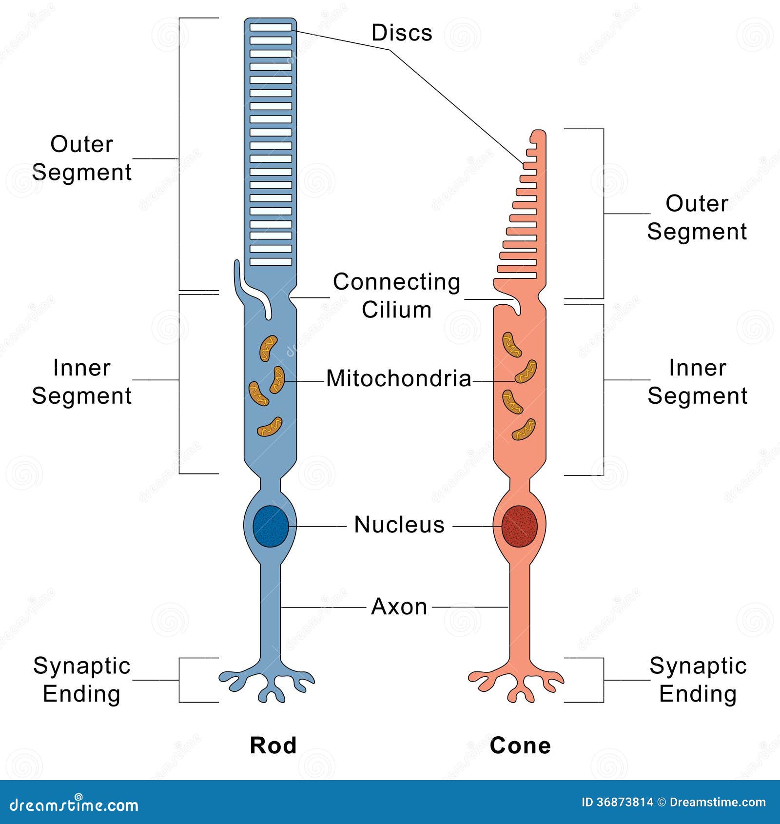
 Huh, like everyone else said, this video didn't illustrate the rods and cones quite right. Cones (and rods) are bipolar neurons. From posterior to anterior a cone goes:outer segment (the "conical" part which contains the photosensitive chemicals, aka the disc), inner segment, cell body (the part that contains the nucleus), axon, axon terminals (aka synaptic ending). Rods have a very similar structure. Here's a diagram to visualize this: http://thumbs.dreamstime.com/z/rod-cone-cells-illustration-human-eye-36873814.jpg(1 vote)
- If the photoreceptors were positioned on the inner side of the retina, there would not need to be a blind spot (optic disk). What is the adaptive quality to the photoreceptor position at the back of the retina?(1 vote)
 That's not true. The optic nerve and the capillaries have to leave SOMEWHERE, so there will always be a spot where there cannot be any photoreceptors!(2 votes)
- 3:00"no axons in the way of the light is that you actually get a higher resolution"
-- does this imply that the increased amount of light hitting the retina increases the resolution of the image?(1 vote)- It kind of does, but I think that's misleading. The benefit of having less axons and cell bodies is that there's a little bit less light scattering, giving a better projection of the outside world onto the retina. This is combined with the massively increased density of photoreceptor cells in the fovea, which is the main contributor to resolution for central vision.(1 vote)
 Many people have commented saying the rods and cones are upside down. What does this mean? Does it mean that the cones are more like inverted cones, with the pointy tip pointing towards the optic nerve, rather than towards the lens? If that is the case, how can rods be upside down, because they're rectangular shaped?(1 vote)
 2:49
2:49
Video transcript
Let's look at how rods and cones
are distributed in the retina. Let me begin by drawing
a very simplified diagram of the eyeball. So in the back of the eye,
we have the optic nerve exiting and going
towards the brain. The back of the eyeball's coated
by a specialized membrane known as the retina. This dimpled portion of the
retina is known as the fovea, and the part of the
retina directly in front of where the optic nerve
exits the back of the eye is actually known
as the blind spot. This is known as the blind
spot because no photo receptors are present in this area. So let's go ahead and
look at the distribution of rods and cones in the eye. So rods are mainly found in
the periphery of the eyeball, so we're going to use this
blue color to represent rods. And they're found mostly in
the periphery of the eye, so up here and a
little bit over here. And as I mentioned, there
are no photo receptors at the blind spot,
because that's where the optic
nerve actually exits the eye, so there's no
photo receptors there. Cones, on the other hand, which
we'll represent in purple, are actually found
throughout the fovea. So they're found in a
really high concentration near the fovea. And there are no cones
at the blind spot, and there are very few
cones kind of sprinkled throughout the rest of the eye. So they're found
kind of sprinkled throughout the
periphery of the eye. So let's go ahead and
do it on the fovea just to kind of make sense of
what we're looking at. So if we zoomed in on the
fovea, what we would see would be the retina,
so it dimples in at the region
where the fovea is. This region from here
to here is the fovea. So let's go ahead and draw
in the rods and cones. So rods are found,
as I mentioned, outside of the fovea, so there
are a whole bunch of rods in the eye specifically
in the periphery. So outside of the fovea, there
are a whole bunch of rods. And there are more
runs over here, and they kind of line the
periphery of the eye and so on. Cones, on the other
hand, are found in a really high
concentration near the fovea. So at the fovea, there are
a whole bunch of cones. And in the periphery
of the eye, there might be a few cones
every now and then. So the reason that the fovea
actually dimples in here is because these photo
receptors are connected to other neurons that
actually send axons through the optic
nerve into the brain. So there are a whole
bunch of neurons over here in this
region, and they all have axons that actually
go to the optic nerve and exit the back of the eye. And so when light enters the
eye and hits the fovea, what it actually looks
like is like this. And so the benefit of having no
axons in the way of the light is that you actually
get a higher resolution, so you actually get more
light is able to hit the cones rather than get
absorbed by these axons. So if I were to enter
this way and hit the periphery of the eye,
what you'd have light entering that actually have to go
through this bundle of axons. And as it's going through, some
of the energy is actually lost, so less light actually hits
rods and cones in the periphery. So at the fovea, you actually
have light directly hitting the cones rather
than having to go through a layer of
axons and neurons. So let's look at this same
picture in another way. Let's go ahead and
look at it graphically. So if I were to draw
a graph, and if I were to say that the 0 point on
the graph-- so let's go ahead and this is the x-axis,
this is the y-axis-- and the 0 point is going
to be where the fovea is. So for you to actually take
the retina-- so in the eyeball the retina's curved like this. If we were to actually
flatten it out, there's a little dimpled region
here that's known as the fovea, and we're going
to set this to 0. And then you can move away, so
this could be five degrees away from the fovea, this could be
10 degrees away from the fovea, this is 15, and so
on on both ends. So that's what we're
going to do here. So this is going to be 5
degrees away from the fovea, this is 10 degrees, and so on. And we're going to
have the same thing on the other side of
the fovea as well. So now on the y-axis,
what we're going to have is receptor density. So this is the number of
receptors found in the retina. So over here we have a
low receptor density, and over here we have a
high receptor density. So as I mentioned, this region
right here is the fovea. And this outside
region from here to here and from here to
here is the periphery. So in the periphery, there's
a really high level of rods. And as we get towards the
fovea, the rods drop in numbers. And then as we start to move
away from the fovea again, there's a really
high number of rods. And we actually reach a region,
which is the blind spot. So there, as I mentioned,
there are no photo receptors where the blind spot is. And as we get to the other
side of the blind spot, there are again photo sectors. And so you kind of get this
type of distribution of rods. Cones have a different
distribution. So cones, there aren't very
many of them in the periphery, but when you get to
the fovea, there's a huge spike in the
number of cones. And as you again move
away from the fovea, the cones actually
drop back down. And then at the blind spot,
there are no photo receptors. And on the other side
of the blind spot, there's a very low number
of photo receptors.